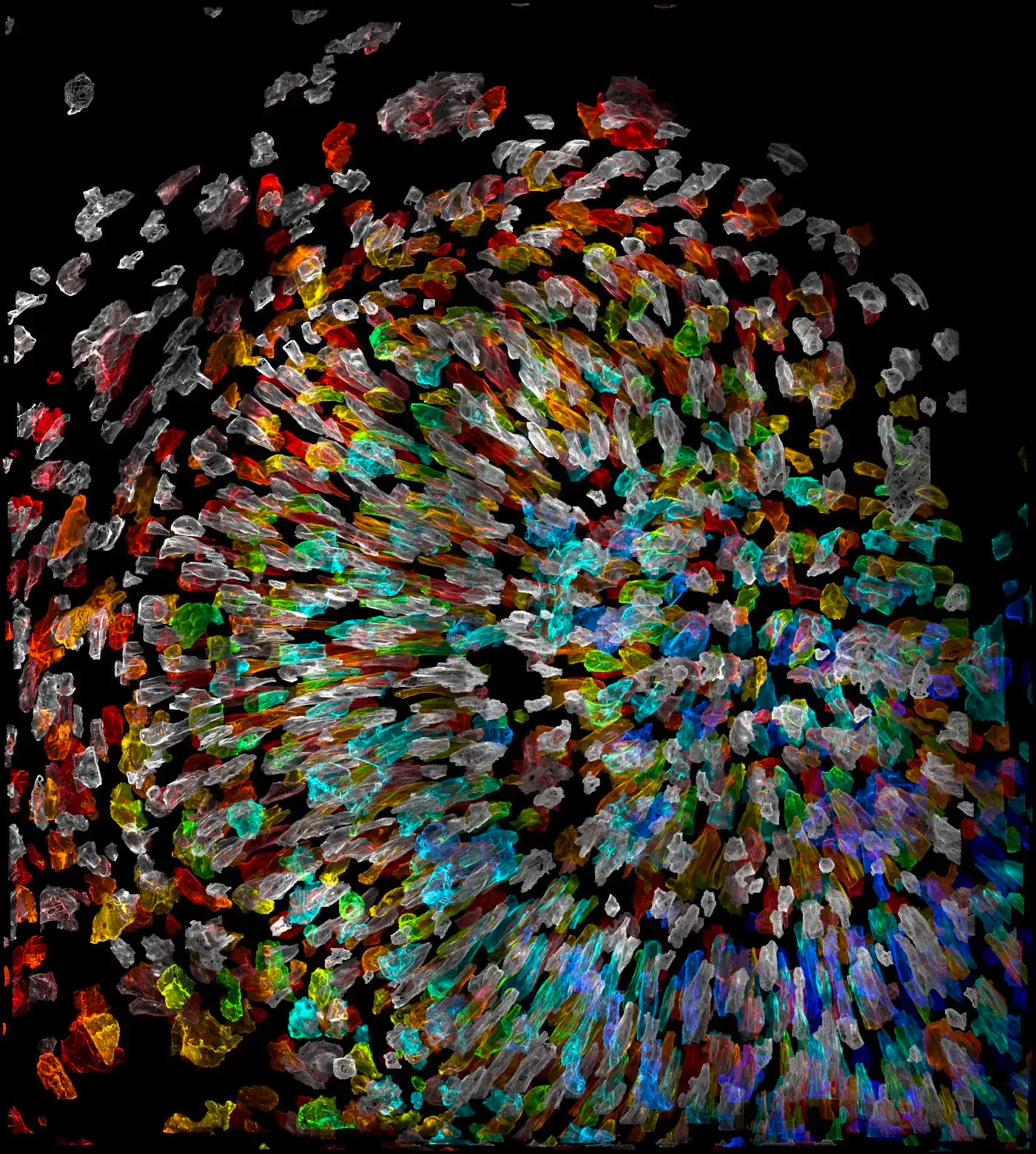
The future of biomedicine is being visualized, quite literally, through the lens of cutting-edge imaging technologies. Scientists are now able to peer deeper into the human body than ever before, observing cells, organs, and entire systems in action. This revolution in visualization is paving the way for groundbreaking discoveries in our understanding of health and disease. The Chan Zuckerberg Initiative’s (CZI) Imaging program is at the forefront of this movement, supporting the development of innovative hardware and open-source software that can capture biological processes across various spatial scales.
This #ImagingTheFuture Week, we spotlight eight remarkable projects led by imaging grantees who are pushing the boundaries of what’s possible. Their work promises to unlock new insights into the inner world of our cells, leading to more effective treatments and preventive measures. Join us as we explore these transformative technologies and their potential to reshape the landscape of healthcare.
Cost-Effective, High-Resolution Correlative Light and Electron Microscopy
Correlative light and electron microscopy (CLEM) is a sophisticated technique that combines the structural preservation of cells with the protein-specific information revealed by light microscopy. Traditionally, CLEM has been limited by its complexity and reliance on specialized expertise and equipment. Lucy Collinson and her team are democratizing CLEM through the VP-CLEM-KIT, a cost-effective pipeline for high-resolution CLEM that can be implemented in standard light microscopy facilities.
“VP-CLEM-KIT will not only enable CLEM research at smaller institutions, but will also have field applications — bringing advanced electron microscopy to more biologists and researchers across the globe.” This innovation broadens access to advanced imaging, empowering researchers worldwide to investigate cellular functions and disease mechanisms with greater precision.
Building a Microscope to View All Information in the Electron Beam
Holger Müller and his team at the University of California, Berkeley, are tackling the challenge of maximizing information extraction from electron microscopy. They are integrating laser phase plate (LPP) technology into a state-of-the-art electron microscope, enabling high-throughput data acquisition and optimizing image quality.
“The microscope will improve the signal-to-noise ratio, allowing researchers to obtain 3D, high-contrast images of proteins in a cell or tissue to visualize in atomic resolution how these molecules work together.” This advancement promises to revolutionize our understanding of molecular interactions and cellular processes.
Arboretum Plugin to Track How Our Cells Are Related
Understanding the interconnectedness of cells is crucial for unraveling complex biological systems. Alan Lowe and his team at University College London have developed Arboretum, a plugin for napari that visualizes cell lineages as a “family tree”.
“Arboretum shows a ‘family tree’ for a given sample of cells, indicating which are alike, and to what degree. The applications for this program go far beyond the cellular level — they can help scientists understand how disease states, samples, or even how entire biological systems are related.” By revealing cellular relationships, Arboretum provides valuable insights into disease progression and biological interactions.
Chip-Scale Light Sheet for High Spatiotemporal Resolution Imaging
Aseema Mohanty and Gokul Upadhyayula are developing a rapidly reconfigurable nanophotonic chip to generate optical lattices for ultra-thin, light sheet-based volumetric imaging of subcellular processes. Their Chip-based Lattice Light sheet Microscope (ChiLL Microscope) will enable flexible, simplified, compact, and low-cost implementation of high-resolution microscopy.
“The ChiLL Microscope’s nanophotonic chip also allows for customization to a researcher’s unique imaging needs. This will expand access to advanced imaging techniques, enabling more researchers to view biology in action at high resolution.” This innovation promises to democratize access to high-resolution imaging, empowering researchers to explore cellular dynamics with unprecedented detail.
HiP-CT Technology Images with Near Cellular Resolution Larger Samples Than Ever Before
Peter Lee and team are expanding the horizons of high-resolution x-ray tomography with Hierarchical Phase-Contrast Tomography (HiP-CT) technology. HiP-CT images larger specimen sizes with the same resolution as small x-ray tomography samples.
“This allows us to image intact human organs at near cellular resolution, providing us with the ability to link microscopic features to macroscopic consequences. This technology has already been used to visualize the impact of COVID-19 on lung health ex vivo, indicating areas of health and damage on a larger and more detailed scale than previously possible and providing clues to what causes Long COVID.” By bridging the gap between microscopic and macroscopic scales, HiP-CT offers invaluable insights into organ-level pathology and disease mechanisms.
Using Deep Learning to Augment Scientists’ Expertise
Réka Hollandi developed the napari-annotatorj plugin as an easy and quick way to add annotations to any 2D image. The technology uses deep learning-based contour suggestions to assist with annotations, with an interface that doesn’t require expertise in computer science.
“Not only does Réka’s plugin help researchers better understand biology at the cellular level, it has the potential to annotate and classify images for a wide variety of fields, from vehicle dash-cam footage to industrial pipeline monitoring.” This plugin streamlines image analysis, making it more accessible and efficient for researchers across various disciplines.
High-Speed Photoacoustic Imaging in Glassfrogs
Junjie Yao and Vladislav Verkhusha have developed a comprehensive toolbox to enhance photoacoustic imaging, breaking the previous resolution limit and enabling never-before-seen depth and clarity of images.
“Their technology was recently applied to better understand how the glassfrog turns itself nearly invisible when sleeping to avoid predators. Using Yao and Verkhusha’s photoacoustic imaging technology, researchers found that glassfrogs store their blood in their liver while sleeping, draining it from areas of the body where it might be visible to predators. Yao and Verkhusha’s advanced imaging technology has enabled discoveries that have exciting potential human applications.” Their enhanced photoacoustic imaging techniques enable non-invasive visualization of biological processes at unprecedented depths and resolution, opening new avenues for understanding physiology and disease.
Enabling a New Type of Microscopy for Ultradeep Imaging
Randy Bartels and team are pushing the bounds of imaging tissues to depths by enabling a new type of microscopy. By measuring a nonlinear distortion operator, they can suppress multiply scattered light that degrades image quality.
“This, in turn, allows high-resolution imaging at significantly greater depths, enabling scientists to see fine details that they have never been able to visualize before.” By overcoming the limitations of light scattering, this technology enables high-resolution imaging deep within tissues, revealing previously inaccessible details of biological structures and processes.
These eight projects represent just a glimpse of the transformative potential of cutting-edge imaging technologies. By pushing the boundaries of what’s possible, these scientists are unlocking new insights into the inner workings of our cells and paving the way for breakthroughs in the prevention, management, and cure of diseases. The Chan Zuckerberg Initiative’s commitment to advancing imaging hardware and software is accelerating this progress, promising a future where we can visualize and understand the complexities of life with unprecedented clarity.
As we continue to #ImagingTheFuture, we can anticipate even more revolutionary tools and techniques that will redefine our understanding of health and disease. The journey into the inner world of our cells has just begun, and the possibilities are limitless.
